Ultrasound technology has become increasingly significant in the medical field, offering non-invasive methods to diagnose various health conditions. One specific area of interest is the use of ultrasound for examining the middle finger. Whether it's related to injuries, joint issues, or other medical concerns, understanding ultrasound middle finger can provide valuable insights into diagnosing and treating conditions effectively.
Ultrasound imaging plays a crucial role in modern medicine, enabling healthcare professionals to visualize internal structures with precision. This article will delve into the importance of ultrasound middle finger, exploring its applications, benefits, and limitations. By the end, you'll have a clear understanding of how this technology impacts patient care.
Whether you're a medical professional, student, or someone curious about this topic, this article aims to provide comprehensive information. We'll cover everything from the basics of ultrasound technology to advanced applications in examining the middle finger. Let's dive in!
Read also:Sssniperwolf Brothers The Untold Story Of Gaming Stardom
Table of Contents
- Introduction to Ultrasound Middle Finger
- Understanding Ultrasound Technology
- Applications of Ultrasound in Middle Finger Examination
- Benefits of Using Ultrasound for Middle Finger
- Limitations of Ultrasound Middle Finger
- Diagnosing Conditions with Ultrasound
- The Procedure for Ultrasound Middle Finger
- Cost Considerations
- Current Research and Developments
- Future of Ultrasound Technology
- Conclusion
Introduction to Ultrasound Middle Finger
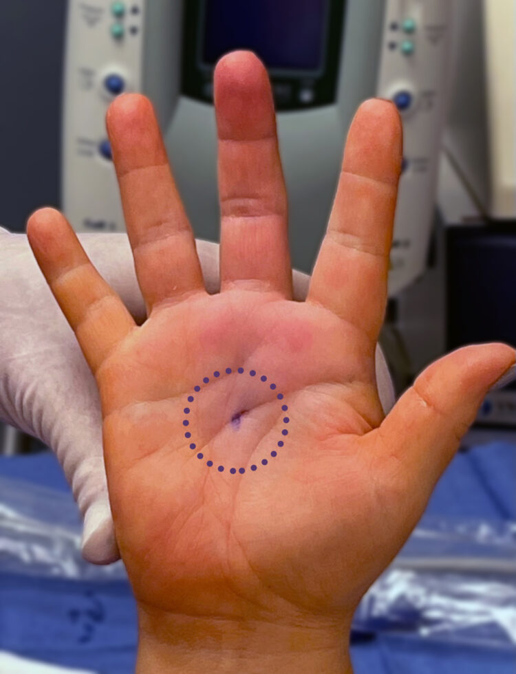
Ultrasound middle finger is a specialized application of ultrasound technology that focuses on examining the middle finger's anatomy and detecting any underlying issues. This technique is particularly useful for diagnosing conditions such as tendon injuries, joint disorders, and soft tissue abnormalities.
Medical professionals often rely on ultrasound imaging to provide a clear picture of the middle finger's internal structures without the need for invasive procedures. By using high-frequency sound waves, ultrasound creates detailed images that help in accurate diagnosis and treatment planning.
In recent years, the use of ultrasound for middle finger examination has gained popularity due to its effectiveness and safety. This section will explore the basics of ultrasound technology and its relevance to middle finger imaging.
Understanding Ultrasound Technology
How Ultrasound Works
Ultrasound technology operates by emitting high-frequency sound waves that bounce off internal structures and return as echoes. These echoes are then converted into visual images by the ultrasound machine. The process is safe, painless, and does not involve radiation, making it an ideal choice for various medical examinations.
Components of an Ultrasound Machine
An ultrasound machine consists of several key components:
- Transducer: The device that emits and receives sound waves.
- Processor: Converts the sound waves into images.
- Display: Shows the resulting images for analysis.
These components work together to produce high-quality images that aid in diagnosing conditions related to the middle finger.
Read also:Travis Scotts Daughter Malu Trevejo The Rising Star In The Spotlight
Applications of Ultrasound in Middle Finger Examination
Ultrasound middle finger has numerous applications in the medical field. Some of the most common uses include:
- Identifying tendon injuries such as tears or inflammation.
- Diagnosing joint disorders like arthritis or synovitis.
- Detecting soft tissue abnormalities such as cysts or tumors.
- Evaluating the effectiveness of treatments over time.
By providing real-time imaging, ultrasound allows healthcare professionals to make informed decisions quickly and accurately.
Benefits of Using Ultrasound for Middle Finger
Non-Invasive and Safe
One of the primary advantages of ultrasound middle finger is its non-invasive nature. Unlike X-rays or CT scans, ultrasound does not involve radiation, making it safe for repeated use. This is especially important for patients requiring regular monitoring.
Cost-Effective
Ultrasound is generally more cost-effective compared to other imaging techniques. It reduces the need for expensive procedures and provides valuable information at a fraction of the cost.
Real-Time Imaging
Ultrasound offers real-time imaging, allowing healthcare professionals to observe dynamic movements and changes in the middle finger's structures. This feature is invaluable for diagnosing conditions that involve motion or function.
Limitations of Ultrasound Middle Finger
While ultrasound middle finger is a powerful diagnostic tool, it does have certain limitations. For example:
- It may not provide detailed images of bone structures compared to X-rays or MRI.
- Obesity or excessive soft tissue can affect image quality.
- Interpretation of results requires skilled professionals with expertise in ultrasound imaging.
Despite these limitations, ultrasound remains a valuable tool in the hands of experienced practitioners.
Diagnosing Conditions with Ultrasound
Ultrasound middle finger plays a crucial role in diagnosing various conditions affecting the finger. Some of the most common conditions include:
Tendon Injuries
Tendon injuries are a frequent cause of pain and discomfort in the middle finger. Ultrasound can identify tears, inflammation, or other abnormalities in the tendons, guiding appropriate treatment options.
Joint Disorders
Conditions such as arthritis or synovitis can significantly impact joint function. Ultrasound imaging helps in detecting early signs of these disorders, allowing for timely intervention.
Soft Tissue Abnormalities
Cysts, tumors, or other soft tissue abnormalities can be accurately identified using ultrasound technology. This aids in determining the best course of action for treatment.
The Procedure for Ultrasound Middle Finger
The process for conducting an ultrasound middle finger examination is straightforward and typically involves the following steps:
- The patient's hand is positioned to ensure optimal imaging of the middle finger.
- A gel is applied to the skin to facilitate the transmission of sound waves.
- The transducer is moved over the area, capturing images from different angles.
- The healthcare professional analyzes the images and provides a diagnosis or recommendations for further testing.
This procedure is generally quick, painless, and well-tolerated by patients.
Cost Considerations
The cost of an ultrasound middle finger examination can vary depending on factors such as location, healthcare provider, and insurance coverage. On average, the procedure is more affordable compared to other imaging techniques like MRI or CT scans.
Insurance often covers ultrasound examinations when deemed medically necessary. Patients are encouraged to check with their providers for specific coverage details.
Current Research and Developments
Ongoing research continues to enhance the capabilities of ultrasound technology. Recent advancements include:
- Improved image resolution for better diagnostic accuracy.
- Integration with artificial intelligence for automated analysis.
- Development of portable ultrasound devices for increased accessibility.
These developments promise to further expand the applications of ultrasound middle finger in the medical field.
Future of Ultrasound Technology
The future of ultrasound technology looks promising, with continued innovations expected to revolutionize medical imaging. As technology advances, ultrasound middle finger examinations will become even more precise, accessible, and cost-effective.
Healthcare professionals and researchers are optimistic about the potential of ultrasound to improve patient outcomes and enhance diagnostic capabilities.
Conclusion
In conclusion, ultrasound middle finger is a vital tool in modern medicine, offering safe, effective, and non-invasive methods for diagnosing conditions related to the middle finger. Its applications span a wide range of medical issues, from tendon injuries to joint disorders and soft tissue abnormalities.
By understanding the benefits, limitations, and procedures involved, patients and healthcare professionals can make informed decisions about using ultrasound for middle finger examinations. We encourage you to share your thoughts or experiences in the comments below, and don't forget to explore other informative articles on our site.